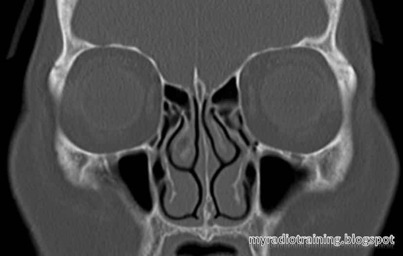
Normal variants in the paranasal sinuses
These are important in radiology report, as gives the endoscopist an idea what to look out for (Surgical planning).
|
Name
|
Location
|
Implication
|
|
||
|
Agger nasi cell
|
Ethmoid aircells.
Located most anteriorly, infront of the cribriform plate where the middle
turbinate attaches
|
If inflamed, patient may experience epiphora
as it is close to the medial canthus
|
|
||
|
Haller cell
(MaxilloEthmoidal cell, Infraorbital cell) |
Aircells located
along the margin of the orbital floor. Inferolateral to the ethmoidal bulla.
|
Presence of these cells may narrow the
infundibulum and/or maxillary sinus ostium. Prone to obstruction and
inflammation of the maxillary sinus.
|
|
||
|
Onodi cell
|
Ethmoid aircells
that extends into the sphenoid bone, located superior to the sphenoid sinus.
|
At risk of intracranial extension of the
endoscope if surgeon (endoscopist) is not aware of the presence of Onodi
cell.
|
|
||
|
Concha bullosa
|
Pneumatized middle
turbinate. These cells usually communicates with the anterior ethmoid
aircells.
|
Large concha bullosa enlarges the turbinate, makes one prone to obstruction/
inflammation.
Concha bullosa also makes endoscopic access
more difficult.
|
|


 |
| What normal variant(s) did you see here? |