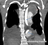
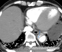
Note: DeBakey system is less commonly used now.
How I remember DeBakey system:
Type I for ONE whole stretch (from ascending to descending), the rest of the types are A and B.
How I remember Stanford's classification:
Type A for Ascending, Type B for Below.
Landmark:
- Left subclavian artery: Distal to this = Descending artery
Important remarks in a report: *remember to evaluate the whole aorta from top down*
- Stanford type A (surgical management) or B (non-surgical management)
- Rupture or not? Presence of haemothorax is a quick and easy sign
- Origin and extent of the dissection
- Presence of aneurysm / Diameter of the aorta
- Identify the false and true lumen (important in interventional management) - this can be identified by tracing the dissection to the end, delayed enhancement in the false lumen
- Identify the complications:
- Involvement of the important arteries: coronary arteries, common carotid arteries, iliac arteries
- Involvement of end organs eg kidneys, intestines, resulting in ischemia / infarction (this is an indication for surgical management)
- Cardiac tamponade as a result of bleed into the pericardium
- Heart failure
- Identify presence of haematoma in the mediastinum, pleura, pericardial or aorta
Associated conditions:
- Connective tissue disorder: Marfan syndrome, Ehlers-Danlos syndrome
- Turner syndrome
- Conditions where the aorta is subjected to high pressure:
- Hypertension
- Aortic coarctation
- Aortic stenosis, Bicuspid aortic valve
- Pregnancy
- Cystic medial necrosis
Differential diagnosis:
- Penetrating atherosclerotic ulcer
- Intramural haematoma (IMH) - also a form of aortic dissection, but without breaching the intima
- Pseudodissection as a result of motion artifact on CT scanning
In any case, remember to pick up the phone and call the surgeons! Happy reporting.


