Tuesday, October 25, 2011

Normal Variant: Cervical Rib


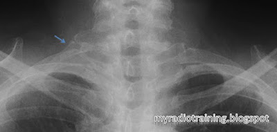
Note the corresponding transverse process points caudally => Cervical origin
Incidental finding of Cervical rib.
Can be unilateral or bilateral.
0.5 - 8% of population*
Usually asymptomatic, but occasionally may give rise to complications.
Think about the anatomical structures that goes through the thoracic outlet, and the attachment of the scalene muscle.

Complications:
- Usually in adulthood
- Thoracic outlet syndrome
- Subclavian artery aneurysm as a result of compression

DDx:
- Hypoplastic 1st rib (rudimentary 1st rib)
- Elongated transverse process of C7

Key to differentiate from the other DDx:
(1) Check transverse process. Cervical transverse process points down, 1st thoracic transverse process points up.
(2) Check presence of joint space between the rib and the transverse process. Absent joint space means it's a elongated transverse process.

Right cervical rib. Note the transverse process pointing down.


*Reference from: Guttentag AR, Salwen JK. Keep your eyes on the ribs: the spectrum of normal variants and diseases that involve the ribs. RadioGraphics 1999;19:1125-1142

Wednesday, October 5, 2011

Case: Aortic Dissection and its classification


 Classification for Aortic Dissection


DeBakey
Stanford


I
A (60%)
Involves both Ascending and Descending aorta


Intimal flap present in both ascending (small circle) and descending (large circle) aorta.
Same patient on Coronal view. Note the peri-aorta haematoma (pink arrow)


II
Involves the Ascending aorta, up to the aortic arch


Intimal flap present only in the ascending aorta.
Same patient in Coronal view.
Intimal flap in Ascending aorta.
Intimal flaps. Don't mistake the aortic valves for intimal flap.

III
B(40%)
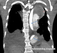
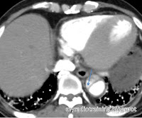
Involves the Descending aorta only.


Involves the Descending aorta only. Note the beak sign (arrow), which signifies the false lumen.
Same patient in Pre-contrast phase.


Note: DeBakey system is less commonly used now.

How I remember DeBakey system: 
Type I for ONE whole stretch (from ascending to descending), the rest of the types are A and B.


How I remember Stanford's classification:
Type A for Ascending, Type B for Below.

Landmark: 
- Left subclavian artery: Distal to this = Descending artery

Important remarks in a report: *remember to evaluate the whole aorta from top down*

  • Stanford type A (surgical management) or B (non-surgical management)
  • Rupture or not? Presence of haemothorax is a quick and easy sign
  • Origin and extent of the dissection
  • Presence of aneurysm / Diameter of the aorta
  • Identify the false and true lumen (important in interventional management) - this can be identified by tracing the dissection to the end, delayed enhancement in the false lumen
  • Identify the complications:
    • Involvement of the important arteries: coronary arteries, common carotid arteries, iliac arteries
    • Involvement of end organs eg kidneys, intestines, resulting in ischemia / infarction (this is an indication for surgical management)
    • Cardiac tamponade as a result of bleed into the pericardium
    • Heart failure
  • Identify presence of haematoma in the mediastinum, pleura, pericardial or aorta

Associated conditions:

  • Connective tissue disorder: Marfan syndrome, Ehlers-Danlos syndrome
  • Turner syndrome
  • Conditions where the aorta is subjected to high pressure:
    • Hypertension
    • Aortic coarctation
    • Aortic stenosis, Bicuspid aortic valve
    • Pregnancy
  • Cystic medial necrosis


Differential diagnosis:
- Penetrating atherosclerotic ulcer
- Intramural haematoma (IMH) - also a form of aortic dissection, but without breaching the intima
- Pseudodissection as a result of motion artifact on CT scanning

In any case, remember to pick up the phone and call the surgeons! Happy reporting.