Thursday, April 16, 2020

Rotator cuff interval - Anatomy


Rotator cuff interval
Triangular shape. Bordered by:
1. Coracoid process
2. Subscapularis muscle/tendon (SSc)
3. Supraspinatus muscle/tendon (SSp)

Houses:
1. (Intra-articular portion of) Long head of biceps tendon (LHBT)
2. Coracohumeral ligament (CHL) (superficial)
     - Variants: absent CHL
     - Originate from postero-lateral of coracoid process
3. Superior glenohumeral ligament (SGHL) (deep)
     - Variants: absent SGHL

Important structures:
1. Medial-pulley-complex (MPC)
     - Medial bundle of CHL + SSc + SGHL
     - Medial to the LHBT
     *Disruption causes subluxation of the LHBT medially

2. Lateral-pulley-complex (LPC)
     - Lateral bundle of CHL + SSp
     - Lateral to the LHBT

3. Rotator cable (part of lateral-pulley-complex)
     - Suspense and stabilise the shoulder joint
     - Thin fibrous band extension from CHL and some of SGHL
     - Traverse deep to SSp and ISp, superficial to joint capsule

4. Capsulo-ligamentous biceps pulley / Superior-complex layer (SCL)
     - CHL + SGHL + Capsule
     - Forms the roof of intra-articular portion of LHBT (proximal to the transverse humeral ligament)

5. Rotator crescent
     - An area between rotator cable and the SSp insertion
     - Poorly vascularised area
     - Gets thinner with age


Reference: https://www.ncbi.nlm.nih.gov/pmc/articles/PMC5568533/

Tuesday, April 14, 2020

Pulled elbow ultrasound

Pulled elbow
aka Nursemaid's elbow
Most common in 1-4 years old

Patho-mechanism
- Traction along the long axis of forearm resulting in subluxation / slippage of annular ligament in between radial head and humerus (capitellum)
- Supinator muscle and annular ligament is slipped over the radial head
- Supinator muscle entraps the radio-humeral joint

Ultrasound examination
Absence of annular ligament in normal position (Most useful sign)
Displaced annular ligament into the radio-humeral joint with enlarged synovial fringe
Abormal shape of the supinator muscle
"J-sign" : supinator muscle and annular ligament trapped in the radio-humeral joint
Look out for fractures as a DDx.


Reference:
https://www.ncbi.nlm.nih.gov/pmc/articles/PMC5052830/#!po=32.1429
Minagawa H. Diagnostic and interventional musculoskeletal ultrasound. J Jpn Orthop Assoc 2012; 86(11): 1057-64.

Monday, April 6, 2020

Elbow PosteroLateral Rotatory Instability Stages

Postero-lateral Rotatory Instability (PLRI) of the elbow

The most common pattern of recurrent elbow instability.
Three stages:

Stage 1:
- Tear / Insufficiency of LUCL
- Postero-lateral subluxation of the radius/ulna on humerus

Stage 2:
- "Perched" dislocation of radius/ulna on humerus
- Coronoid process perched under the tochlea
- Disruption of anterior and posterior joint capsule
- Disruption of the LUCL

Stage 3:
- Complete dislocation of elbow radius/ulna on humerus
- Coronoid process dislocated, positioned behind humerus.
- LUCL, RCL, joint capsule disruption
- MCL disruption
     - Stage 3A: Posterior bundle disrupted ; Anterior bundle intact
     - Stage 3B: Posterior and Anterior bundles disrupted
     - Stage 3C: Disrupted ligaments and tendons on both medial and lateral aspects


Reference:
https://www.ncbi.nlm.nih.gov/pmc/articles/PMC6447645/#!po=73.8636
https://www.ncbi.nlm.nih.gov/pmc/articles/PMC5443138/

Tuesday, March 31, 2020

Dallas classification for discography

Dallas classification

A classification system to assess the degree of annular tear/fissuring
*The term "tear" is not recommended in imaging reporting according to 2014 consensus of the North American Spine Society, American Society of Spine Radiology & American Society of Neuroradiology

Assess via CT Discogram following Discography.
Dallas classification evolves from the initial 4 grades up to 7 grades over time
*Grade 5 annular fissure can be determined during Discography

Grade 0:
- Normal contained nucleus pulposus

Grade 1:
- Contrast leak into the inner 1/3 of annulus

Grade 2:
- Contrast leak into the inner 1/3 & middle 1/3 of annulus

Grade 3:
- Contrast leak into the inner, middle and outer 1/3 of annulus (ie entire thickness of annulus), less than 30% circumference

Grade 4:
- Contrast leak to circumferentially more than 30% of the annulus

Grade 5:
- Contrast leak into epidural space

Grade 6:
- Disc sequestration

Grade 7:
- Diffuse annular tear in disc degeneration


Reference:
https://www.ncbi.nlm.nih.gov/pmc/articles/PMC3097593/#!po=45.0000

Ramsay-Hunt Syndrome

AKA Herpes zoster oticus

Aetiology agent:
- Herpes zoster virus
Latent (Reactivation) infection at geniculate ganglion of virus from past infection (chicken pox)

Structure affected: 
- Facial nerve (CN VII)
- +/- Vestibulocochlear nerve (CN VIII)

MRI:
- Contrast enhancement of the CN VII canalicular segment. Normally do not enhance in this area.
- Asymmetric contrast enhancement of the geniculate ganglia labyrinthine, tympanic and/or mastoid.
- +/- Vestibular & Cochlear nerve contrast enhancement.
- +/- Pontine colliculus facials contrast enhancement.

Symptoms:
Classic triad
- Vesicles along the facial nerve distribution (ear, EAC, face, palate, tongue) AND/OR
- Ipsilateral hemi-facial weakness / paralysis AND/OR
- Ear (otic) pain
Others:
- Tinnitus, sensorineural hearing loss, vertigo (vestibulocochlear nerve CNVIII involvement)
- Unable to close eye (ipsilateral)
- Altered / Loss of taste perception
- Dry mouth & eyes

Complication:
- Facial paralysis (can be permanent)
- Hearing loss (can be permanent)
- Post-herpetic neuralgia
- Meningo-encephalitis

Rx: EARLY TREATMENT!!
- Antiviral medications
- Short term high dose Steroids
- Benzodiazepine
- Analgesia
- Eye-moisturiser

Hallux valgus

Angle between longitudinal axis of 1st metatarsal bone and 1st proximal phalanx >15 degrees deviated laterally.

Associated with
- Foot pronation
- Flat foot

Atrophy of the intrinsic plantar muscles:
- Abductor hallucis (AbdH)
- Flexor hallucis brevis (FHB)
* Not observed in Flexor digitorum brevis (FDB)

Thickening of Plantar fascia:
- Anterior / Middle / Posterior Plantar fascia

Complication:
- Bunion formation
- Progress to Hallux rigidus

Tuesday, March 24, 2020

Mayfield classification

Mayfield classification

Classification for carpal bone instability (peri-lunate injuries)

Mechanism of injury:
- Fall on out-stretched hand (FOOSH)
- Injuries always start from Radial side to Ulnar side, therefore Type 1 -> 4 = injuries progressively from Radial to Ulnar side
- A combination of 3 pressure load (listed below), causes rotatory force around the waist of capitate.

  1. Carpus extension (the principal load)
  2. Wrist ulnar deviation
  3. Intercarpal supination

Important ligaments in perilunate injuries
Purple: Radio-scapho-capitate ligament
Blue: Radio-triquetral ligament
Yellow: Ulno-triquetral ligament

Imaging:
Radiographs are insensitive except for frank perilunate dislocation / lunate dislocation
Clues:

  1. Scapho-lunate dissociation 
    1. Scapho-lunate interval 2-4mm: Suspicious ; >4mm: Definite on AP view
    2. Scapho-lunate angle >63 degree on Lateral view
  2. Scaphoid bone fracture
  3. Radial styloid fracture
  4. Luno-triquetral dissociation
    1. Scaphoid angle <30 degree:="" identify="" li="" means="" no="" on="" only.="" other="" radiograph="" suspicious="" to="">
  5. Triquetrum fracture
  6. Capitate fracture
  7. Gilula's arch disruption / step deformity
  8. "Piece of pie" sign for perilunate dislocation / lunate dislocation
  9. "Spilt tea-cup" sign for lunate dislocation
CT better at depicting avulsion injury, step deformity
MRI / MR Arthrography better at depicting bone marrow oedema (trabecula fracture in instance of trauma), ligamentous integrity


Type 1:

*Area of disruption: Scapho-lunate articulation
- Extension + Ulnar deviation => Radio-scapho-capitate ligament tear/avulsion
- Further force causes => Scaphoid  fracture / Scapho-lunate ligament tear

Mayfield type 1: Scapho-lunate articulation disruption


Type 2:

*Area of disruption: Scapho-lunate + Capito-lunate articulations
- Type 1 injuries plus:
- Capito-lunate ligament tear/avulsion OR Capitate fracture

Mayfield type 2: Scapholunate & Capitolunate articulation disruptions


Type 3:

*Area of disruption: Scapho-lunate + Capito-lunate + Luno-triquetral articulations
- Type 1 + Type2 plus:
- Lunotriquetral ligament tear/avulsion OR Triquetrum fracture
- Resulting in peri-lunate dissociation ie Dissociation of lunate from (1) Scaphoid (radial side) ; (2) Capitate (distal articulation) ; (3) Triquetrum (Ulnar side).
- +/- Peri-lunate dislocation ensues ie Lunate is still articulating normally with radius, whereas the rest of the carpal bones are dislocated dorsally.

Mayfield type 3: Scapholunate, Capitolunate & Lunotriquetral articulation disruptions


Type 4:

*Area of disruption: Scapho-lunate + Capito-lunate + Luno-triquetral + Radio-lunate articulations
- Radio-lunate ligament tear AND
- Relocation of the carpal bones (except Lunate) back to normal position AND
- Lunate is tilted and displaced to volar aspect, through the space of Poirier.

Mayfield type 4: All 4 articulations of the lunate bone disrupted


Reference: https://doi.org/10.1016/j.crad.2019.10.0160009-9260/

Monday, March 23, 2020

Breast nodule below 25 years old - Maxwell criteria


Current recommendation (UK, Ireland) is discharge WITHOUT biopsy or follow-up for:
- Women <25yo and="" p="">- Breast nodule typical of fibroadenoma on Ultrasound examination


Maxwell criteria for non-biopsy of solid masses in <25yo b="" female=""> (Maxwell & Pearson*)

Clinical features:
1. No known risk factors
2. Mass/nodule not rapidly enlarging
3. Smooth discrete mobile nodule / Impalpable

Sonographic features:
1. Well-defined, ovoid shape
2. Flat lesion, ie Height < Width
3. <3cm dimension="" greatest="" in="" p="">4. Smooth outline / Gently lobulated (2-3 lobulations) (NO microlobulations!)
5. Homogeneously isoechoic or slight hyperechoic, solid
6. Thin echogenic pseudocapsule
7. No calcification.
8. No posterior acoustic shadowing.


Reference:
* Maxwell AJ, Pearson JM. Criteria for the safe avoidance of needle sampling in young women with solid breast masses. Clin Radiol 2010;65:218-22

Monday, March 16, 2020

ACNES

Abdominal Cutaneous Nerve Entrapment Syndrome (ACNES)

One of the causes for chronic abdominal pain. Can lead to central sensitisation of the pain
Pain from the abdominal wall - hyperalgesia / allodynia / hyperaesthesia
Often points to lateral border of rectus abdominis

Entrapment of the cutaneous nerve due to:
(1) Sharp turning of the nerve through layers (neurovascular channels) of abdominal wall muscles
(2) Surgical scar tissue entrapment on the nerve (eg Abdominal wall hernia surgery)
(3) Injury to the nerve from surgical incision

Anatomy:
- Abdominal wall cutaneous nerves originate from ventral rami of T7 - T12
- 3 branches: Posterior branch ; Lateral branch ; Anterior branch
- Recall the landmark area:
     - T10 at umbilicus level
- The nerves traverse between Internal Oblique Muscle (IOM) & Transverse Abdominalis (TA)
- The nerves continue anteriorly until behind the rectus abdominis.
- The anterior branch (most common to give rise to symptom): Opening / 'Sharp turn' of the nerve approximately at 0.5-1cm medial to the linea semilunaris (from posterior rectus sheath through the rectus abdominis into the skin, via a fibrous neurovascular channel)


Risk factors:
- Female
- Pregnancy (causes stretching of the muscle wall)
- Previous abdominal surgery
- Sports activity (that requires abdominal muscle contraction)

Clinical features:
- Well localised pain, often unilateral
- Neuropathic pain
- +/- Valleix phenomenon: Retrograde (circumferential) radiation of the pain (DDx Thoracic radiculopathy)
- Pain on movement (ie contraction of wall muscle => compression of nerve)
- Lying down may relief OR aggravate the pain

Examination:
- Carnett's sign
- Hover sign
- No pain on deep palpation (positive tenderness would indicate deep organ pathology)
- Pinch test

Ultrasound-guided injection/hydrodissection (Diagnostic & Therapeutic)
- Aim: Posterior rectus sheath
- 22-25G needle, LA+Steroid / 5% Dextrose water / Pulsed-RF / 50% Alcohol (less used nowadays)
- Scar-related ACNES: Hydrodissect the scar formation area with 5% Dextrose water
- Be mindful of (1) secondary nerve injury (2) the deeper structure - peritoneum
- Some may require multiple injections over a period of time

Other Rx:
- Rectus muscle stretching exercise
- Avoidance of triggering factor (eg certain exercise)
- Scar-related ACNES: Wall exercise to maintain normal gliding muscle movement
- Hot / Cold application
- Topical LA / Capsaicin cream

**T12/L1 nerve involvement may present with pain in scrotum / vulva
**Long list of DDx depending on level of involvement eg costochondritis
**Lateral branch and posterior branch can give rise to symptoms as well, but at different location. (less prone due to oblique orientation of the exiting nerve)

Reference: 
https://www.ncbi.nlm.nih.gov/pmc/articles/PMC6220638/
https://academic.oup.com/bjaed/article/15/2/60/248606


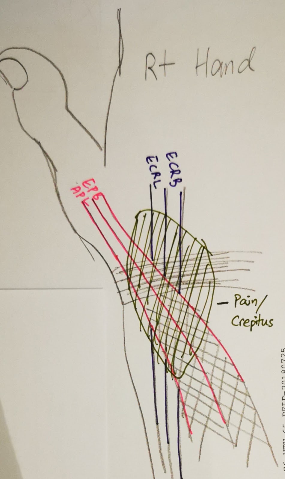
Radial intersection syndrome

(Radial) Intersection Syndrome
An overuse syndrome

Area of intersection between compartment 1 and 2 tendons


SYMPTOMS:
- Radial aspect of wrist & forearm pain, a/w local swelling (inflammation)


EXAMINATION:
- Friction/Crepitus at distal dorso-radial forearm upon active/passive wrist motion (where the tendons intersect)
- Weak and/or pain on wrist & thumb movement
- Tender over radial tubercle and lateral aspect of the anatomical snuff box


ETIOLOGY / RISK FACTORS:
- Tenosynovitis of the extensor muscles - (Dorsal compartment 2) ECRL, ECRB, (Dorsal compartment 1) APL, EPB
- Friction between the tendons of Compartment 1 & Compartment 2 where it intersects in the distal forearm dorso-radially.
- Overuse of wrist extensor muscles, tenosynovitis of the 2nd compartment tendons
- Repeated wrist extension-flexion
- More common in woman, 30-50s
- Athlete: eg Rowers


DDx:
- de Quervain's tenosynovitis (Check Finkelstein test)
- 1st Carpo-metacarpal joint osteoarthritis (CMC OA) (Check thumb grinding maneuvre)
- EPL tendinitis
- Wartenberg syndrome (Superficial sensory Radial nerve neuropathy) (Check Tinel sign)


MANAGEMENT:
- Rest (give 3/52)
- Physical therapy (stretching & strengthening)
- Anti-inflammatory medications
- Splinting - 15-20deg wrist extension thumb splint. (gradually wean off)
- Modification of triggering activity
- Injection & Hydrodissection
- Surgery (last resort)

Reference:
- https://www.jabfm.org/content/30/4/547

Sunday, October 13, 2013

Ultrasound of the spine for dysraphism

To look for occult spinal dysraphism

Clinically features suggestive for occult spinal dysraphism:
- Sacral dimple
- Tuft of hair
- Vascular patch / haemangioma
- Overlying skin appendage / skin tags
- Other anomalies which maybe associated with tethered cord eg imperforate anus, lower limb neurology

When to do / When not to do:
- Avoid performing if patient <2/52 old (often not enough CSF). Can do if high clinical probability.
- 2/52 to 4-6/12
- Not ideal if >6/12 old as spinous processes starts to ossify, obscuring the view

Preparation:
Patient relaxed eg just finished feeding, sleeping
Position: Prone or lying on side
High resolution linear probe

What to look for / document:
Sagittal view: (through the cartilaginous spinous processes)

1. Level of conus: L2/L3 disk or above, may be lower if pre-term
- If 'low lying' in pre-term, followup scan at 40wk after corrected age.
- Low lying cord may imply cord tethering
- Count the vertebral levels (reference point):
-- from below: sacrum or 1st coccygeal segment
-- from above: last rib
- may be supplemented with lateral spine radiograph to aid in counting in difficult cases, marker can be placed at the level of conus termination

2. Morphology of conus: Normally tapers down.

3. Normal pulsating motion of the spinal cord
- Absence in tethering

4. Dependent position of spinal cord (difficult to ascertain if not enough CSF as in the case of performing in patient below 2/52 old)
- Normal: Lie on anterior 1/3 or halfway in the canal.

5. Filum terminale thickness
- Normal </=2mm

Transverse view:

6. Spinal vertebral dysraphism (posterior elements)

Developmental Dysplastic Hip

DDH:
- Acetabular dysplasia AND/OR ligamentous laxity, resulting in recurrent hip subluxation / dislocation
- Left hip affected more than Right hip

Risk factors for DDH:
1. Breech presentation
2. Female (?estrogen contributing to ligamentous laxity?)
3. Firstborn
4. Family Hx
5. Oligohydramnios , Multiple gestation - abnormal posture due to crowding in-utero

Clinical features:
1. Asymmetric thigh skin folds
2. Shortened leg
3. Decreased hip ROM when Flexed and ABducted
4. Positive Ortolani and Barlow signs

Ultrasound used to assess the hips - the cartilaginous components can be seen. ie not ossified yet

Contraindications to US Hips:
1. Generally avoid if patient <3-4/52 old, as the ligaments are normally lax. Usually done at 6/52 and after.
2. Not suitable after 6mo. Femoral head ossifies, starts around 2-3mo.
Radiographs are a better modality of choice when the bone ossifies (>12mo) - AP radiographs, not frog leg view.

Ultrasound:
Done when patient is relaxed. eg. whilst feeding
Use high-resolution linear probe.

1. Coronal view
Hip slightly flexed. (~15-20degrees)
These structures should be seen, in addition to the femoral head, greater trochanter, acetabular roof.
a. Ossified ilium is horizontally orientated
b. Triradiate cartilage
c. Labrum

Look for:
1. Morphology of the hip joint
2. Coverage of femoral head using the straight ilium line (Position)
3. Alpha angle

2. Transverse view
Hip flexed. (90 degrees)
Look for: Hip joint stability
Visualisation of the hip joint whilst performing
- Barlow maneuvre
- If dislocatin/subluxation present, perform Ortolani maneuvre

Graf system (US hip for DDH introduced by Graf - Orthopaedic Surgeon)
- To grade severity of DDH and appropriate management

Graf system

Acetabular rim
Labrum
Ossified ilium line
Alpha angle

Type I
-
Normal angular
Normal
Crosses >50% of femoral head
>60
Normal
Type IIa

Age: <3mo
Rounded
Normal
Crosses <50% of femoral head, but not displaced
50-59
Physiologic immaturity.
Repeat scan in 6-8/52

Type IIb
Age: >3mo
Same finding as IIa
Refer orthopaedic
Type IIc
Same finding as IIa
Everted / horizontal position
Same finding as IIa
43-49
Type IIIa
Normal acetabular roof cartilage
Flattened
-
Displaced (<50%)
<43
Refer orthopaedic
Type IIIb
Abnormalechogenic acetabular roof cartilage
Same finding as IIIa
Type IV
-
Flattened

Complete displacement
<43
Urgent orthopaedic referral

Radiographs:
1. Asymmetric hip joints
2. Asymmetric femoral epiphysis ossification: As a complication of DDH where there is delayed ossification
3. Femoral head crossing the Perkin's line laterally
4. Acetabular angle >30degree
5. Disruption of Shenton's line

Hilgenreiner's line:
Transverse line touching the triradiate cartilage superiorly and bilaterally on AP pelvis view

Perkin's line:
Line perpendicular to Hilgenreiner's line, touching lateral margin of the ossified acetabular roof on AP pelvis view.

Acetabular angle:
Angle between Hilgenreiner's line and the acetabular roof.
Normal: <30degree

Shenton's line:
Smooth curve lining the inferior margin of the superior pubic ramus and the medial margin of the proximal femur.

Putti's triad:
1. Femoral head displace superolaterally
2. Small femoral epiphysis
3. Acetabular angle > 35degrees

Monday, July 22, 2013

Cotswold modification of Ann-Arbor Classification for Hodgkin lymphoma

For staging and prognosis of Hodgkin lymphoma.

Stage I
1 or 2 contiguous regions of lymph nodes on same side of diaphragm
Stage II
> 2 contiguous regions of lymph nodes on same side of diaphragm
Stage III
Both sides of diaphragm
Stage IV
Organ involvement*
X
Bulky disease >10cm
E
Extranodal involvement – used in conjunction with stage I-III
A/B
A: Absence of symptoms / B: Presence of constitutional symptoms


*Thymus and Spleen are lymphoid organ, thus included in the ‘lymph node’ involvement. They are not considered as ‘organ’ in this context.

Sarcoidosis

Sarcoidosis
Idiopathic. Granulomatous disease (non-caseating).
Systemic disease involving the lymph nodes, lungs, liver, heart etc.
Lung is the most commonly involved organ.

Clinical features:
- 3rd or 4th decades
- More in African american
- 70% have increased serum ACE - An indicator of the granuloma burden on the body

Nuclear Medicine:
- Gallium (Ga 67 scintigraphy) avid during active phase. No uptake when in remission.
Degree of uptake corresponds to degree of severity
Confirms diagnosis of sarcoidosis
Differentiate active alveolitis from inactive fibrosis
Signs:
  1. Lambda sign: Hilar and Right paratracheal nodal uptake
  2. Diffuse pulmonary uptake
  3. Panda sign: Lacrimal and salivary glands inflammation

- PET scan: FDG avid during active phase. SUV can be >2.5

Acute Sarcoidosis
aka LÓ§fgren syndrome
Clinical features:
- Fever, malaise, arthralgia +/- parotitis / uveitis
- Erythema nodosum
- Serum ACE level often increased
- Erythema nodosum
Has bilateral hilar lymphadenopathy, but absence of lung changes

Lungs:
Garland’s triad (1-2-3 sign)
Lambda sign on Gallium scan
  1. Bilateral hilar lymphadenopathy
  2. Right paratracheal lymphadenopathy
CXR:
-          Reticulonodular opacities, bilateral upper lobe dominant
-          Symmetric bilateral hilar, paratracheal, aortopulmonary window lymphadenopathy

HRCT:
-          Lymphadenopathy
-     1-5mm nodules in centrilobular and subpleural distribution
-     Upper lobe and peribronchovascular distribution
-     Irregular interlobular septal thickening
-     Architectural distortion
-          Advanced: Interstitial fibrosis- Peripheral honeycombing, Bullae
-     Some cases (rare): Alveolar sarcoidosis = Peripheral consolidation + Mediastinal and Bilat.hilar lymphadenopathy

Radiographic staging:
-     Stage 0: Normal CXR
-          Stage I: disease limited to mediastinal nodes
-          Stage II: involvement of lung and lymph nodes
-          Stage III: involvement of lung only
-          Stage IV: lung fibrosis

DDx:
- When advanced, can mimic progressive massive fibrosis (PMF)


Cardiac
Result in:
- Arrhythmia
- Ventricular aneurysms
- Sudden death

Head & Neck:
Involvement of glandular tissues, muscles, nerves.
1. Optic nerve and EOM hypertrophy and enhancement - uveitis
2. Parotid glands and lacrimal glands exhibits heterogeneous bilateral symmetrical enlargement - chronic dacroadenitis, parotitis

Gallium scan: "Panda sign" where there is increased uptake of the parotid and lacrimal glands (Looks like the whole panda sitting on the person's neck / replacing the head)

Neurological system:
Neurosarcoidosis, 5% of sarcoidosis patient
Usually lungs would have been involved already.
Granulomatous infiltration of the perineural tissue
Involvement of nerves, optic chiasm, hypothalamus, infundibulum, IAM
Presenting with cranial nerve deficits

Imaging:
Exhibit contrast smooth / nodular enhancements along the involved nerves, thickened leptomeninges, pachymeninges
Also enhancement in the perivascular space, ependyma
5% have solitary extra-axial mass

Complication: Hydrocephalus

Gastrointestinal:
Spleen:
Splenomegaly
Present as splenic granulomas, +/- calcification
If present = advanced stage of Sarcoidosis.
Spontaneous splenic rupture (rare)

Lymph nodes:
Mesenteric nodes


Musculoskeletal:
Affect small bones of hands and feet
Reticulated trabecular pattern (lace-like)
Cyst-like lesions
Destruction of terminal phalanges
Diffuse sclerosis of the vertebral bodies
Osteolysis in skull

Monday, April 22, 2013

Bonescan lesions

3 +/- 1 phase Bone scan
1st phase: Arterial blood flow; immediately after injection
2nd phase: Blood pool (tissue phase) 10min after injection
3rd phase: Delayed phase (Skeletal phase) 2-5hr after injection
4th phase: 24hr delayed phase if renal insufficiency and as a result suboptimal image on 3rd phase

Standard whole body bone scan: Acquire at delayed phase (skeletal phase)
Mechanism: Radiotracer adsorbed on hydroxyapatite matrix of the bone, where the mineral phase is active.

Superscan:
Increased radiotracer uptake of bone in relative to the soft tissue background and (almost) absent renal activity.
Causes:

  1. Widespread metastasis in: Breast / Lung / Prostate CA, +/- Lymphoma +/- Bladder CA
  2. Metabolic bone disease: Renal osteodystrophy, Osteomalacia, Hyperparathyroidism
  3. Myelofibrosis, Systemic mastocytosis


Hot lesions:
  1. Fibrous dysplasia
  2. Paget's disease
  3. Brown tumors
  4. ABC (because it is very vascular)
  5. Osteoid osteoma
  6. Chondroblastoma
  7. Metastasis (Osteoblastic activities ie Sclerotic eg Prostate CA mets)
  8. Joint diseases
  9. Dental infection
  10. Acute fracture (after 24 - 48hr)

3 phase positive lesions: (lesions that causes bone remodelling)
  1. Osteomyelitis
  2. Healing fracture
  3. Orthopaedic implant
  4. Neuropathic osteoarthropathy

Normal scan: (no increased or decreased uptake)
  1. Acute fracture (acute phase)
  2. Bone cyst, bone island, exostoses
  3. Multiple myeloma ** there for this condition is monitored by skeletal survey radiograph**
  4. Osteoporosis
Cold lesions:
  1. Radiotherapy
  2. Avascular lesion
  3. Multiple myeloma 
  4. Haemangioma
  5. Advanced carcinoma
Varying lesions
Mixed lytic/sclerotic metastasis (Breast, Lung etc) may show hot or cold (with hot margins).

Monday, December 10, 2012

Enostosis - Bone island

Commonly known as bone island

Mature cortical bone within the medulla
Due to failure of resorption during endochondral ossification
Asymptomatic

Features:
CT/Radiograph: 
- Spiculated margin - blends with the surrounding trabecular bone
- Dense sclerotic focus
MR:
- All sequences: Very low SI
Scintigraphy
- Typically no activity. Some may have increased activity

DDx:
- Dense osteoid osteoma
- Osteoblastoma

PVNS

Pigmented villonodular synovitis
- Unknown aetiology
- Synovial proliferation disorder
- intracellular Haemosiderin deposition (thus "Pigmented")

Monoarticular
Age 30-40s

Benign proliferation of synovium in the joint or in bursa/tendon sheath +/- hemorrhagic effusion
If occurs in the tendon sheath => then it is known as Giant Cell Tumor of the tendon sheath

Clinical features:
- Insidious onset
- Monoarticular arthropathy
- Swelling
- Stiffness
- Pain

Pathology:
- Hyperplastic synovium
- Lipid-laden foam cells, histiocytes, giant cells, haemosiderin deposits

Distribution:
- Knee > Hip > Ankle > Shoulder

Radiograph:
- Dense effusion
- Dense soft tissue mass (due to haemosiderin)
- Bone erosion
- Large subchondral cysts
- Preserved joint space
- Typically normal in the early stage

MRI:
- Joint effusion
- Focal / diffuse synovial thickening: Low signal T1w and T2w, and blooming artifact on GRE (due to hemosiderin deposition)

DDx:
- (repeated) Haemarthrosis such as Haemophilics
- Septic arthritis
- Articular amyloid deposition (similar in MRI appearance)

Rx:
- Synovectomy (~50% with local recurrence)

Wednesday, October 17, 2012

Bosniak classification for Renal Cyst

Applicable to US, CT or MR imaging.


Features
Management

Bosniak I
Anechoic cyst
Thin walled
No septa, calcification, solid components
No contrast enhancement
No intervention required
Class 1 Renal cyst

Bosniak II
Hairline-thin septa
Fine calcification / Short thick calcification
No contrast enhancement
No intervention required
Class 2 Renal Cyst

Class 2. Notice the faint thin septa?
Also a smaller simple cyst in the lower pole.

Bosniak IIF
Multiple hairline-thin septae +/- minimal thickening
Minimal wall thickening
Thick / Nodular calcification
No soft tissue component
Needs follow-up

Bosniak III
Thick wall / septa, irregular / smooth
Needs surgical intervention
Class 3. Notice the thicker septa and larger size of the cyst?
Class 3 Renal Cyst

Bosniak IV
Bosniak III + soft tissue component which enhances.
Clearly malignant

Thursday, December 29, 2011

Psoriatic arthritis

Psoriatic arthritis

Polyarticular arthritis
Skin disease: Psoriasis (Skin rash)
Chronic inflammatory
40yo, Caucasian
?Factors involved: Genetic, Immune system, Environmental
HLA-B27 +ve in 60%

Hand:
- Most commonly involved skeleton
- Interphalangeal joint (PIJ, DIJ)
- 'Fluffy periostitis'
- Bony proliferation*
- Joint erosion from articular margin* to centrally.
- Tapering of one end, saucerisation of the other end of the articular surface: Pencil-in-cup deformity
- Preserved bony mineralisation (no osteopenia), ivory phalanx
- Ankylosis
- Diffuse soft tissue swelling: Sausage digit

SIJs:
- Asymmetric sacroiliitis, sclerosis, large erosion
- Ankylosis is UNcommon

Vertebra:
- More commonly involved in MALE
- Asymmetric
- Atlanto-axial subluxation
- Squarring of vertebrae
- Enthesitis: comma-shaped ossification, paravertebral (parasyndesmophytes)
- Thoracolumbar region

Others:
- MTPJ
- TA insertion of the Calcaneum: Fluffy periostitis & Erosion

*HALLMARKS for Psoriatic arthritis

DDx:
- Ankylosing spondylitis (Ossification less bulky, symmetric sacroiliitis)
- Reactive arthritis (Genitourinary symptoms)
- Hyperparathyroidism (similarity: presence of large SIJ erosion)

Monday, November 7, 2011

Modic changes in MRI


Modic classification

Use for different endplate marrow degenerative changes (which is postulated to result from same pathological process, but at different stages) in lumbar spine MR imaging.

Modic degenerative changes often accompanies disk degenerative disease (DDD)


The Modic change can be of mix type 1 and 2, type 2 and 3.

Modic type
T1w
T2w
Pathology
1
(oedema)
Hypointense


Hyperintense

Modic1. Note also disk dessication.

Oedema & Inflammation
2
(fat)
Hyperintense


Iso/Slight Hyperintense

Modic2. Note also disk dessication.

Marrow ischaemia, thus red marrow converts to fatty yellow marrow
3
(sclerosis)
Hypointense


Hypointense

Modic3. Note also disk dessication.

Subchondral sclerosis

Radiograph of the same patient with Modic 3 degeneration. Note sclerosis (arrow).


Note:
Disciitis can mimic Modic 1 degenerative change, when the adjacent bone marrows from the inflamed/infected disk is inflamed

Look at 3 things:
(1) Whether the intervertebral disk shows corresponding degenerative change (disk dessication) or shows inflammed (disciitis)

(2) Whether the bone or adjacent structures points to infection eg bone erosion, abscess formation

(3) Last but not least, correlate clinically - Lab changes, Clinical history


Endplates
Intervertebral disk
Others
Modic 1
Low T1w, High T2w
Iso/Low T2w

Usually dessicated (modic change accompanies disk degenerative disease)

Clinically non-toxic
Diskiitis
Same as above, but with features of diskiitis ie bone erosion
High T2w

Inflammed disk
Presence of inflammation / abscess in adjacent structures (eg paraspinal, epidural abscess)

Clinical and laboratory findings of infection