Tuesday, March 31, 2020

Hallux valgus

Angle between longitudinal axis of 1st metatarsal bone and 1st proximal phalanx >15 degrees deviated laterally.

Associated with
- Foot pronation
- Flat foot

Atrophy of the intrinsic plantar muscles:
- Abductor hallucis (AbdH)
- Flexor hallucis brevis (FHB)
* Not observed in Flexor digitorum brevis (FDB)

Thickening of Plantar fascia:
- Anterior / Middle / Posterior Plantar fascia

Complication:
- Bunion formation
- Progress to Hallux rigidus

Tuesday, March 24, 2020

Mayfield classification

Mayfield classification

Classification for carpal bone instability (peri-lunate injuries)

Mechanism of injury:
- Fall on out-stretched hand (FOOSH)
- Injuries always start from Radial side to Ulnar side, therefore Type 1 -> 4 = injuries progressively from Radial to Ulnar side
- A combination of 3 pressure load (listed below), causes rotatory force around the waist of capitate.

  1. Carpus extension (the principal load)
  2. Wrist ulnar deviation
  3. Intercarpal supination

Important ligaments in perilunate injuries
Purple: Radio-scapho-capitate ligament
Blue: Radio-triquetral ligament
Yellow: Ulno-triquetral ligament

Imaging:
Radiographs are insensitive except for frank perilunate dislocation / lunate dislocation
Clues:

  1. Scapho-lunate dissociation 
    1. Scapho-lunate interval 2-4mm: Suspicious ; >4mm: Definite on AP view
    2. Scapho-lunate angle >63 degree on Lateral view
  2. Scaphoid bone fracture
  3. Radial styloid fracture
  4. Luno-triquetral dissociation
    1. Scaphoid angle <30 degree:="" identify="" li="" means="" no="" on="" only.="" other="" radiograph="" suspicious="" to="">
  5. Triquetrum fracture
  6. Capitate fracture
  7. Gilula's arch disruption / step deformity
  8. "Piece of pie" sign for perilunate dislocation / lunate dislocation
  9. "Spilt tea-cup" sign for lunate dislocation
CT better at depicting avulsion injury, step deformity
MRI / MR Arthrography better at depicting bone marrow oedema (trabecula fracture in instance of trauma), ligamentous integrity


Type 1:

*Area of disruption: Scapho-lunate articulation
- Extension + Ulnar deviation => Radio-scapho-capitate ligament tear/avulsion
- Further force causes => Scaphoid  fracture / Scapho-lunate ligament tear

Mayfield type 1: Scapho-lunate articulation disruption


Type 2:

*Area of disruption: Scapho-lunate + Capito-lunate articulations
- Type 1 injuries plus:
- Capito-lunate ligament tear/avulsion OR Capitate fracture

Mayfield type 2: Scapholunate & Capitolunate articulation disruptions


Type 3:

*Area of disruption: Scapho-lunate + Capito-lunate + Luno-triquetral articulations
- Type 1 + Type2 plus:
- Lunotriquetral ligament tear/avulsion OR Triquetrum fracture
- Resulting in peri-lunate dissociation ie Dissociation of lunate from (1) Scaphoid (radial side) ; (2) Capitate (distal articulation) ; (3) Triquetrum (Ulnar side).
- +/- Peri-lunate dislocation ensues ie Lunate is still articulating normally with radius, whereas the rest of the carpal bones are dislocated dorsally.

Mayfield type 3: Scapholunate, Capitolunate & Lunotriquetral articulation disruptions


Type 4:

*Area of disruption: Scapho-lunate + Capito-lunate + Luno-triquetral + Radio-lunate articulations
- Radio-lunate ligament tear AND
- Relocation of the carpal bones (except Lunate) back to normal position AND
- Lunate is tilted and displaced to volar aspect, through the space of Poirier.

Mayfield type 4: All 4 articulations of the lunate bone disrupted


Reference: https://doi.org/10.1016/j.crad.2019.10.0160009-9260/

Monday, March 23, 2020

Breast nodule below 25 years old - Maxwell criteria


Current recommendation (UK, Ireland) is discharge WITHOUT biopsy or follow-up for:
- Women <25yo and="" p="">- Breast nodule typical of fibroadenoma on Ultrasound examination


Maxwell criteria for non-biopsy of solid masses in <25yo b="" female=""> (Maxwell & Pearson*)

Clinical features:
1. No known risk factors
2. Mass/nodule not rapidly enlarging
3. Smooth discrete mobile nodule / Impalpable

Sonographic features:
1. Well-defined, ovoid shape
2. Flat lesion, ie Height < Width
3. <3cm dimension="" greatest="" in="" p="">4. Smooth outline / Gently lobulated (2-3 lobulations) (NO microlobulations!)
5. Homogeneously isoechoic or slight hyperechoic, solid
6. Thin echogenic pseudocapsule
7. No calcification.
8. No posterior acoustic shadowing.


Reference:
* Maxwell AJ, Pearson JM. Criteria for the safe avoidance of needle sampling in young women with solid breast masses. Clin Radiol 2010;65:218-22

Monday, March 16, 2020

ACNES

Abdominal Cutaneous Nerve Entrapment Syndrome (ACNES)

One of the causes for chronic abdominal pain. Can lead to central sensitisation of the pain
Pain from the abdominal wall - hyperalgesia / allodynia / hyperaesthesia
Often points to lateral border of rectus abdominis

Entrapment of the cutaneous nerve due to:
(1) Sharp turning of the nerve through layers (neurovascular channels) of abdominal wall muscles
(2) Surgical scar tissue entrapment on the nerve (eg Abdominal wall hernia surgery)
(3) Injury to the nerve from surgical incision

Anatomy:
- Abdominal wall cutaneous nerves originate from ventral rami of T7 - T12
- 3 branches: Posterior branch ; Lateral branch ; Anterior branch
- Recall the landmark area:
     - T10 at umbilicus level
- The nerves traverse between Internal Oblique Muscle (IOM) & Transverse Abdominalis (TA)
- The nerves continue anteriorly until behind the rectus abdominis.
- The anterior branch (most common to give rise to symptom): Opening / 'Sharp turn' of the nerve approximately at 0.5-1cm medial to the linea semilunaris (from posterior rectus sheath through the rectus abdominis into the skin, via a fibrous neurovascular channel)


Risk factors:
- Female
- Pregnancy (causes stretching of the muscle wall)
- Previous abdominal surgery
- Sports activity (that requires abdominal muscle contraction)

Clinical features:
- Well localised pain, often unilateral
- Neuropathic pain
- +/- Valleix phenomenon: Retrograde (circumferential) radiation of the pain (DDx Thoracic radiculopathy)
- Pain on movement (ie contraction of wall muscle => compression of nerve)
- Lying down may relief OR aggravate the pain

Examination:
- Carnett's sign
- Hover sign
- No pain on deep palpation (positive tenderness would indicate deep organ pathology)
- Pinch test

Ultrasound-guided injection/hydrodissection (Diagnostic & Therapeutic)
- Aim: Posterior rectus sheath
- 22-25G needle, LA+Steroid / 5% Dextrose water / Pulsed-RF / 50% Alcohol (less used nowadays)
- Scar-related ACNES: Hydrodissect the scar formation area with 5% Dextrose water
- Be mindful of (1) secondary nerve injury (2) the deeper structure - peritoneum
- Some may require multiple injections over a period of time

Other Rx:
- Rectus muscle stretching exercise
- Avoidance of triggering factor (eg certain exercise)
- Scar-related ACNES: Wall exercise to maintain normal gliding muscle movement
- Hot / Cold application
- Topical LA / Capsaicin cream

**T12/L1 nerve involvement may present with pain in scrotum / vulva
**Long list of DDx depending on level of involvement eg costochondritis
**Lateral branch and posterior branch can give rise to symptoms as well, but at different location. (less prone due to oblique orientation of the exiting nerve)

Reference: 
https://www.ncbi.nlm.nih.gov/pmc/articles/PMC6220638/
https://academic.oup.com/bjaed/article/15/2/60/248606


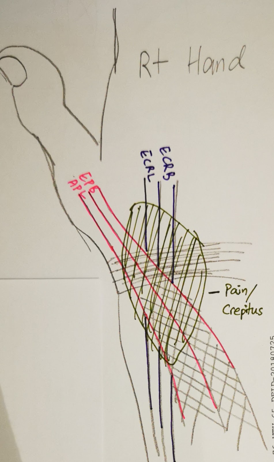
Radial intersection syndrome

(Radial) Intersection Syndrome
An overuse syndrome

Area of intersection between compartment 1 and 2 tendons


SYMPTOMS:
- Radial aspect of wrist & forearm pain, a/w local swelling (inflammation)


EXAMINATION:
- Friction/Crepitus at distal dorso-radial forearm upon active/passive wrist motion (where the tendons intersect)
- Weak and/or pain on wrist & thumb movement
- Tender over radial tubercle and lateral aspect of the anatomical snuff box


ETIOLOGY / RISK FACTORS:
- Tenosynovitis of the extensor muscles - (Dorsal compartment 2) ECRL, ECRB, (Dorsal compartment 1) APL, EPB
- Friction between the tendons of Compartment 1 & Compartment 2 where it intersects in the distal forearm dorso-radially.
- Overuse of wrist extensor muscles, tenosynovitis of the 2nd compartment tendons
- Repeated wrist extension-flexion
- More common in woman, 30-50s
- Athlete: eg Rowers


DDx:
- de Quervain's tenosynovitis (Check Finkelstein test)
- 1st Carpo-metacarpal joint osteoarthritis (CMC OA) (Check thumb grinding maneuvre)
- EPL tendinitis
- Wartenberg syndrome (Superficial sensory Radial nerve neuropathy) (Check Tinel sign)


MANAGEMENT:
- Rest (give 3/52)
- Physical therapy (stretching & strengthening)
- Anti-inflammatory medications
- Splinting - 15-20deg wrist extension thumb splint. (gradually wean off)
- Modification of triggering activity
- Injection & Hydrodissection
- Surgery (last resort)

Reference:
- https://www.jabfm.org/content/30/4/547

Sunday, October 13, 2013

Ultrasound of the spine for dysraphism

To look for occult spinal dysraphism

Clinically features suggestive for occult spinal dysraphism:
- Sacral dimple
- Tuft of hair
- Vascular patch / haemangioma
- Overlying skin appendage / skin tags
- Other anomalies which maybe associated with tethered cord eg imperforate anus, lower limb neurology

When to do / When not to do:
- Avoid performing if patient <2/52 old (often not enough CSF). Can do if high clinical probability.
- 2/52 to 4-6/12
- Not ideal if >6/12 old as spinous processes starts to ossify, obscuring the view

Preparation:
Patient relaxed eg just finished feeding, sleeping
Position: Prone or lying on side
High resolution linear probe

What to look for / document:
Sagittal view: (through the cartilaginous spinous processes)

1. Level of conus: L2/L3 disk or above, may be lower if pre-term
- If 'low lying' in pre-term, followup scan at 40wk after corrected age.
- Low lying cord may imply cord tethering
- Count the vertebral levels (reference point):
-- from below: sacrum or 1st coccygeal segment
-- from above: last rib
- may be supplemented with lateral spine radiograph to aid in counting in difficult cases, marker can be placed at the level of conus termination

2. Morphology of conus: Normally tapers down.

3. Normal pulsating motion of the spinal cord
- Absence in tethering

4. Dependent position of spinal cord (difficult to ascertain if not enough CSF as in the case of performing in patient below 2/52 old)
- Normal: Lie on anterior 1/3 or halfway in the canal.

5. Filum terminale thickness
- Normal </=2mm

Transverse view:

6. Spinal vertebral dysraphism (posterior elements)

Developmental Dysplastic Hip

DDH:
- Acetabular dysplasia AND/OR ligamentous laxity, resulting in recurrent hip subluxation / dislocation
- Left hip affected more than Right hip

Risk factors for DDH:
1. Breech presentation
2. Female (?estrogen contributing to ligamentous laxity?)
3. Firstborn
4. Family Hx
5. Oligohydramnios , Multiple gestation - abnormal posture due to crowding in-utero

Clinical features:
1. Asymmetric thigh skin folds
2. Shortened leg
3. Decreased hip ROM when Flexed and ABducted
4. Positive Ortolani and Barlow signs

Ultrasound used to assess the hips - the cartilaginous components can be seen. ie not ossified yet

Contraindications to US Hips:
1. Generally avoid if patient <3-4/52 old, as the ligaments are normally lax. Usually done at 6/52 and after.
2. Not suitable after 6mo. Femoral head ossifies, starts around 2-3mo.
Radiographs are a better modality of choice when the bone ossifies (>12mo) - AP radiographs, not frog leg view.

Ultrasound:
Done when patient is relaxed. eg. whilst feeding
Use high-resolution linear probe.

1. Coronal view
Hip slightly flexed. (~15-20degrees)
These structures should be seen, in addition to the femoral head, greater trochanter, acetabular roof.
a. Ossified ilium is horizontally orientated
b. Triradiate cartilage
c. Labrum

Look for:
1. Morphology of the hip joint
2. Coverage of femoral head using the straight ilium line (Position)
3. Alpha angle

2. Transverse view
Hip flexed. (90 degrees)
Look for: Hip joint stability
Visualisation of the hip joint whilst performing
- Barlow maneuvre
- If dislocatin/subluxation present, perform Ortolani maneuvre

Graf system (US hip for DDH introduced by Graf - Orthopaedic Surgeon)
- To grade severity of DDH and appropriate management

Graf system

Acetabular rim
Labrum
Ossified ilium line
Alpha angle

Type I
-
Normal angular
Normal
Crosses >50% of femoral head
>60
Normal
Type IIa

Age: <3mo
Rounded
Normal
Crosses <50% of femoral head, but not displaced
50-59
Physiologic immaturity.
Repeat scan in 6-8/52

Type IIb
Age: >3mo
Same finding as IIa
Refer orthopaedic
Type IIc
Same finding as IIa
Everted / horizontal position
Same finding as IIa
43-49
Type IIIa
Normal acetabular roof cartilage
Flattened
-
Displaced (<50%)
<43
Refer orthopaedic
Type IIIb
Abnormalechogenic acetabular roof cartilage
Same finding as IIIa
Type IV
-
Flattened

Complete displacement
<43
Urgent orthopaedic referral

Radiographs:
1. Asymmetric hip joints
2. Asymmetric femoral epiphysis ossification: As a complication of DDH where there is delayed ossification
3. Femoral head crossing the Perkin's line laterally
4. Acetabular angle >30degree
5. Disruption of Shenton's line

Hilgenreiner's line:
Transverse line touching the triradiate cartilage superiorly and bilaterally on AP pelvis view

Perkin's line:
Line perpendicular to Hilgenreiner's line, touching lateral margin of the ossified acetabular roof on AP pelvis view.

Acetabular angle:
Angle between Hilgenreiner's line and the acetabular roof.
Normal: <30degree

Shenton's line:
Smooth curve lining the inferior margin of the superior pubic ramus and the medial margin of the proximal femur.

Putti's triad:
1. Femoral head displace superolaterally
2. Small femoral epiphysis
3. Acetabular angle > 35degrees